自動化細胞活性分析平台
Automated Living Cell Assay Platform
A4-高通量共軛焦顯微影像系統
ImageXpress Micro Confocal
ImageXpress Micro Confocal High-Content Imaging System

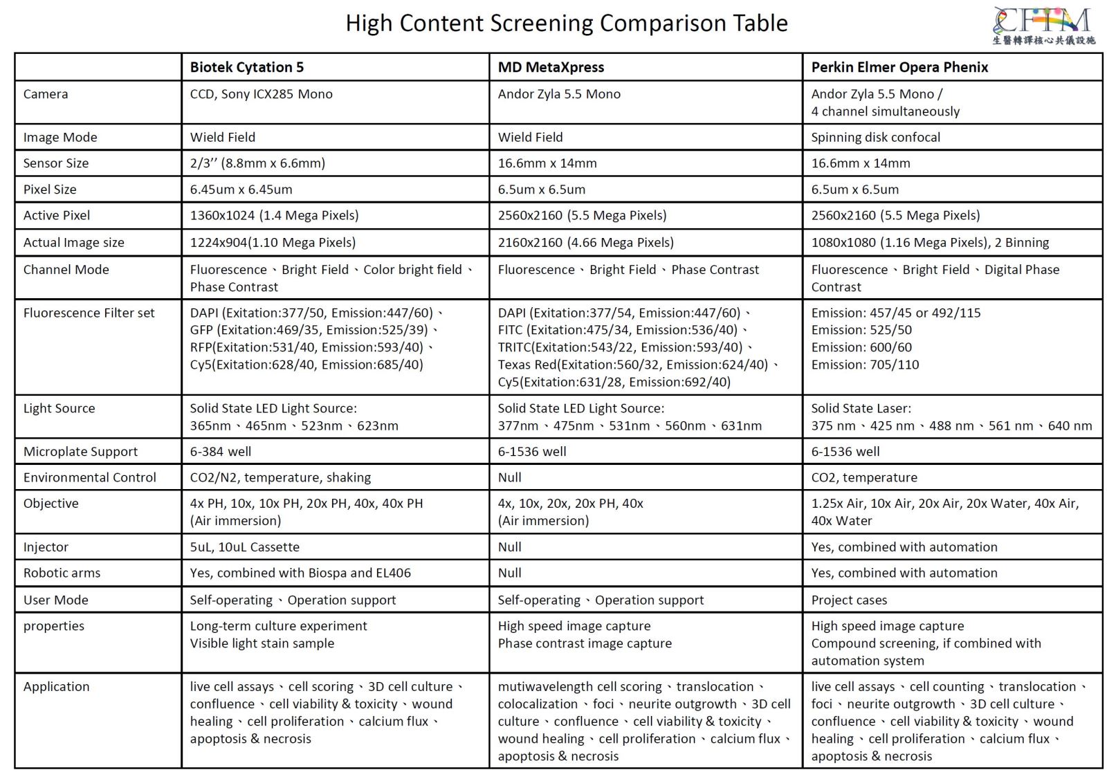
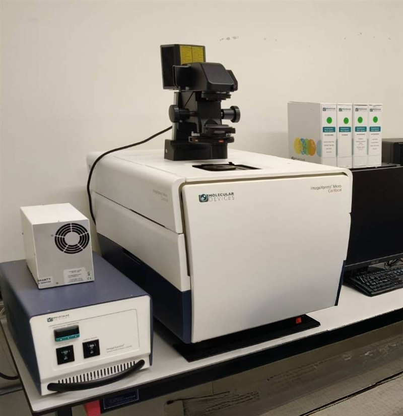
儀器描述
MD ImageXpress Micro Confocal High-Content Imaging System 為高內涵影像分析系統(High-Content Imaging System),具備高通量、多孔盤式細胞影像擷取及定量分析能力,可自動化於短時間內進行大量樣品之螢光影像擷取與分析,並針對特定細胞族群及多項細胞參數進行定量分析。
本系統整合廣域螢光(Widefield)與 spinning disk 共軛焦(Spinning Disk Confocal)成像功能,可進行 Z 軸影像掃描、3D 影像擷取及 3D 定量分析,並支援固定細胞與活細胞影像實驗。適用於細胞凋亡、細胞毒性、細胞型態及其他多參數高內涵細胞分析等研究。
主要特點
- 高通量、高內涵細胞影像擷取與定量分析
- 支援 6–1536 well 多孔盤及玻片影像擷取
- 支援固定、活細胞長時間細胞影像拍攝與分析
- Z 軸影像掃描及 3D 影像擷取
- 3D 影像定量分析
- 自動化影像擷取與多參數分析
- 支援廣域場及共軛焦成像模式切換
- 雙自動對焦系統,包含雷射自動對焦(Laser Autofocus)及影像自動對焦(Image Autofocus)
儀器規格
多孔盤支援:
支援 6–1536 well 多孔盤及玻片。
光源:
固態螢光光源(Solid State Light Source)。
螢光濾鏡:
配置 DAPI、FITC、Cy3、Texas Red 及 Cy5 等 5 組螢光濾鏡組。
自動對焦系統:
具備雷射自動對焦(Laser Autofocus)及影像自動對焦(Image Autofocus)雙重對焦功能,可提升大量樣品影像擷取之效率及穩定性。
共軛焦成像系統:
配置 spinning disk 共軛焦(Spinning Disk Confocal)模組,提供 60 µm pinhole 及 50 µm slit 兩種共軛焦選項,並可依實驗需求切換共軛焦與廣域場成像模式。
物鏡:
- 4× Plan Apo LAMBDA,NA 0.20
- 10× Plan Apo,NA 0.45
- 20× Ph1 S Plan Fluor ELWD ADM,NA 0.45
- 40× S PLAN FLUOR ELWD,NA 0.60
相機:
Andor Zyla 4.2。
環境控制模組:
具備活細胞實驗所需之環境控制功能,支援 6–1536 well 多孔盤,可控制溫度範圍為室溫至 40°C,並配置濕度維持水槽及氣體流速控制器,以提供適合活細胞長時間觀察之實驗環境。
控制及分析軟體:
MetaXpress Software,支援儀器控制、影像擷取及影像分析;另提供離線影像分析功能,可進行影像定量及多參數分析。
使用注意事項
使用 10×以上物鏡進行影像擷取時,應搭配薄底或玻璃底培養盤,以確保影像品質及成像效果。
放置地點
國家生技研究園區A棟5樓A502a室
管理人員
陸韻翔 專員
電子信箱:lululoh@gate.sinica.edu.tw
電話:02-7750-5556
Instrument Description
The MD ImageXpress Micro Confocal High-Content Imaging System is a high-content imaging system designed for high-throughput, automated acquisition and quantitative analysis of cellular images. The system enables rapid fluorescence imaging of large numbers of samples in multiwell plates and supports quantitative analysis of specific cell populations and multiple cellular parameters.
The system integrates both widefield fluorescence and spinning-disk confocal imaging capabilities, allowing users to perform Z-stack imaging, 3D image acquisition, and quantitative 3D image analysis. It supports both fixed-cell and live-cell imaging applications and is suitable for a wide range of high-content cellular assays, including apoptosis, cytotoxicity, cell morphology, and multiparametric cellular analysis.
Key Features
- High-throughput and high-content cellular imaging and quantitative analysis
- Supports 6- to 1536-well microplates and slide imaging
- Imaging of fixed cells
- Live-cell imaging capabilities
- Z-axis scanning and Z-stack image acquisition
- 3D image acquisition and quantitative analysis
- Automated image acquisition and multiparametric analysis
- Switchable widefield and spinning-disk confocal imaging modes
- Dual autofocus systems, including laser autofocus and image autofocus
Instrument Specifications
Plate Compatibility:
Supports 6- to 1536-well microplates and slides.
Light Source:
Solid-state light source.
Fluorescence Filters:
Five fluorescence filter sets: DAPI, FITC, Cy3, Texas Red, and Cy5.
Autofocus System:
Equipped with dual autofocus systems, including laser autofocus and image autofocus, to provide efficient and stable focusing during automated high-throughput image acquisition.
Confocal Imaging System:
Equipped with a spinning-disk confocal module with two selectable confocal options, including a 60 µm pinhole and 50 µm slit. The system can be switched between confocal and widefield imaging modes according to experimental requirements.
Objectives:
- 4× Plan Apo LAMBDA, NA 0.20
- 10× Plan Apo, NA 0.45
- 20× Ph1 S Plan Fluor ELWD ADM, NA 0.45
- 40× S PLAN FLUOR ELWD, NA 0.60
Camera:
Andor Zyla 4.2.
Environmental Control Module:
The system is equipped with an environmental control module for live-cell imaging. It supports 6- to 1536-well microplates and provides temperature control from room temperature to 40°C, together with a humidity-maintaining water reservoir and gas-flow controller to provide a suitable environment for long-term live-cell imaging.
Control and Analysis Software:
MetaXpress Software is used for instrument control, image acquisition, and image analysis. Offline image analysis is also available for quantitative and multiparametric image analysis.
Important Note
When using 10× or higher magnification objectives, thin-bottom or glass-bottom culture plates should be used to ensure optimal imaging quality and performance.
Facility location
National Biotechnology Research Park Building A, 5th Floor, room A502a
Manager
陸韻翔 技術專員
Email:lululoh@gate.sinica.edu.tw
TEL:02-7750-5556
